Magnetic Sample Modulation AFM
Selective modulation of magnetic domains can be accomplished with AFM imaging by applying an AC current to a solenoid placed under the sample stage. Magnetic nanomaterials will vibrate according to the amplitude and strength of the applied field. A nonmagnetic tip that is scanned in continuous contact with the sample can be used to detect the vibration of magnetic nanomaterials.
![]()
A soft, nonmagnetic cantilever is used to detect motion and vibrational response rather than directly measuring magnetic forces. The information acquired from MSM images include the distribution of individual magnetic domains, which respond to the flux of an AC generated magnetic field, as well as spectra of the characteristic resonance frequencies of the vibrating nanomaterials.
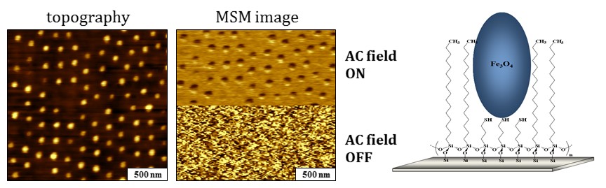 Iron oxide deposits on nanopatterns of 3-mercaptopropyltrimethoxysiloxane. When the
field is turned off half-way through the scan, the phase and amplitude channels show
no distinguishable features of the sample. [Li, J.-R.; Lewandowski, B. R.; Xu, S.;
Garno, J. C. Development of a New Hybrid AFM Imaging Mode for Selective Mapping of
Magnetic Nanomaterials, Anal. Chem. 2009, 81, 4792 .]
Iron oxide deposits on nanopatterns of 3-mercaptopropyltrimethoxysiloxane. When the
field is turned off half-way through the scan, the phase and amplitude channels show
no distinguishable features of the sample. [Li, J.-R.; Lewandowski, B. R.; Xu, S.;
Garno, J. C. Development of a New Hybrid AFM Imaging Mode for Selective Mapping of
Magnetic Nanomaterials, Anal. Chem. 2009, 81, 4792 .]
Essentially, MSM is a variation of force modulation AFM, however with selectivity for magnetic nanomaterials. The periodic motion of the sample vibration can be tracked by changes in the deflection of the AFM tip as it touches a vibrating area of the sample. Changes in the phase angle and amplitude as the tip interacts with the vibrating nanoparticles are plotted as a function of tip position to create MSM phase and amplitude images. The mechanical motion of the sample will produce contrast exclusively for vibrating domains responding to the flux of the magnetic field.
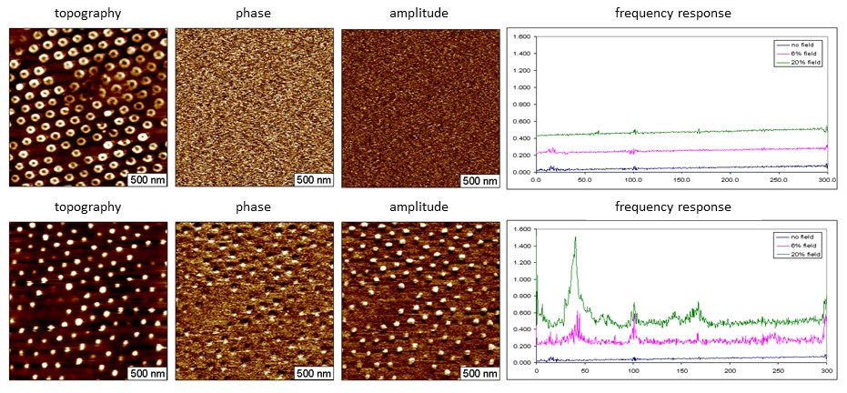
MSM experiment showing the tip response for a sample that does not contain iron (top row) and the same sample after depositing iron on the organosilane nanorings (bottom row). The MSM phase and amplitude frames map the locations of vibrating iron oxide deposits.
Images obtained with MSM can visualize how magnetic nanoparticles or metalloproteins respond to the flux of an applied AC electromagnetic field. Results with MSM demonstrate new possibilities for AFM imaging of magnetic nanomaterials such as ferritin, with exceptional sensitivity. Ferritin is a spherical iron storage protein with a hollow shell enclosing a cavity containing iron atoms. The ferrimagnetic core of ferritin is weakly paramagnetic. Particle lithography was used to create nanopatterns of ferritin for MSM studies. Individual ferritin molecules were sensitively detected, in areas in between the rings. The mechanical motion of the sample can be sensitively detected by a scanning AFM tip.
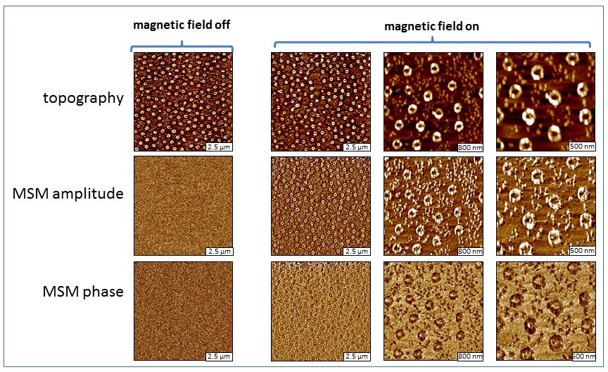
Images of ferritin ring structures acquired with MSM, the magnification is increased from left-to-right. When the electromagnetic field is off, there are no distinguishable features in the amplitude and phase frames (left). When the field is turned on, the areas of ferritin rings are revealed. Individual proteins are detectable in areas between the rings. [Daniels, S. L.; Ngunjiri, J. N.; Garno, J. C. Investigation of the magnetic properties of ferritin by AFM imaging with magnetic sample modulation. Analytical & Bioanalytical Chemistry, 2009, 394 (1), 215-223. ]