BioAFM
Protein patterning is important for emerging nanoscale biological and medical applications, such as for ultrasensitive bioassays. Particle lithography is a practical, highly reproducible method for patterning proteins on surfaces. Proteins can be attached securely to the surface, forming nanopatterns with a measured thickness of a single layer. Periodic arrays of nanostructures offer a valuable test platform for studies of molecular reactions for surface-based bioassays at the molecular level. Changes in the morphology of protein nanostructures can be monitored in situ after chemical agents, nanoparticles or other small molecules are introduced to protein nanopatterns. Well-defined protein arrays offer precise reproducible surface features for multiple measurements at the nanoscale. By performing experiments in aqueous buffers, studies of protein-DNA interactions and antigen–antibody binding can be accomplished by capturing images of the evolution of surface changes during biochemical reactions. The particle templates used for preparing test platforms can be scaled to ever smaller dimensions, for studies of size-dependent properties at tens of nanometers, approaching single-molecule detection.
Nanostructures of Ferritin Prepared with Particle Lithography
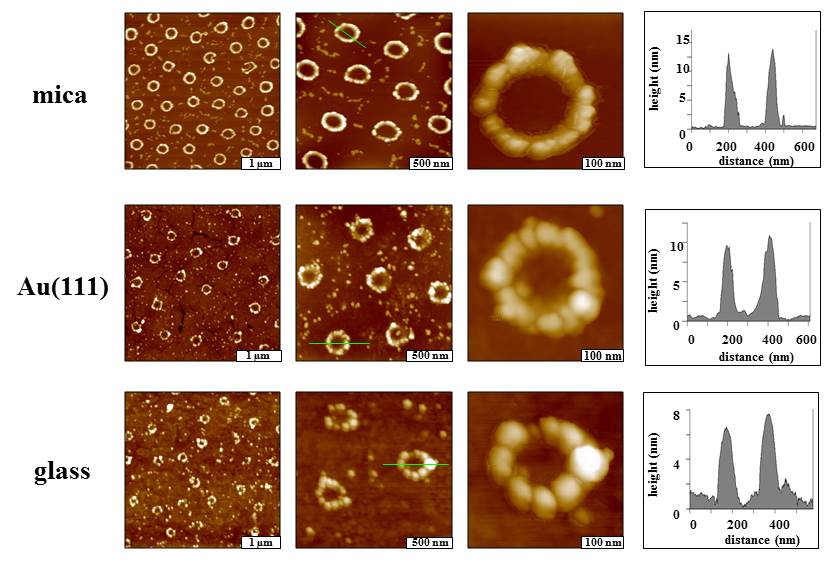
The arrangement of rings of ferritin prepared on several substrates that are commonly used for binding proteins viewed with successive zoom-in topography images. Ferritin is a protein that stores iron, releasing it as needed. The shape of ferritin is nearly spherical and it has a diameter of 10–12 nm. Individual proteins can be resolved in the AFM topography frames.
[Ngunjiri, J. N.; Daniels, S. L.; Li, J.-R.; Serem, W. K.; Garno, J. C. Controlling the surface coverage and arrangement of proteins using particle lithography. (invited) Nanomedicine, 2008, 3(4), 529-541.]
Spatially Selective Binding of Green Fluorescent Protein
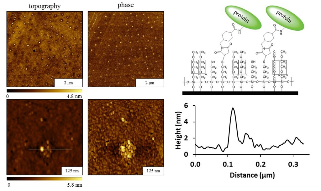
Nanopatterns of (3-mercaptopropyl)trimethoxysilane (MPTMS) surrounded by a methoxy terminated matrix were prepared with particle lithography as a surface template. The sulfhydryl groups of the MPTMS nanopatterns were activated with a sulfosuccinimidyl-4-(N-maleimidomethyl)cyclohexane-1-carboxylate linker. The activated regions of MPTMS provided sites for selective binding of green fluorescent protein.
[Highland, Zachary L.; Garno, J. C. Spatially selective binding of green fluorescent protein on designed organosilane nanopatterns prepared with particle lithography. Biointerphases, 2017, 12(2), 02C402.]
Selective Binding of Fibrinogen on Organosilane Nanopatterns
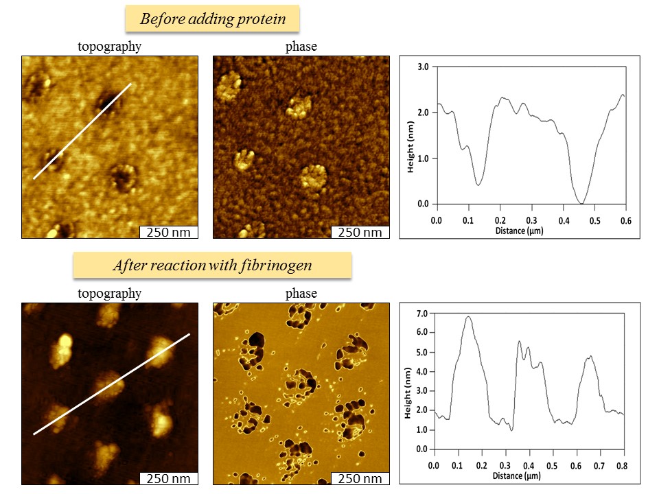
Particle lithography was applied for patterning fibrinogen, a plasma protein that has a major role in the clotting cascade for blood coagulation and wound healing. Surface nanopatterns of mercaptosilanes were designed as sites for the attachment of fibrinogen within a protein-resistant matrix of 2-[methoxy(polyethyleneoxy)propyl] trichlorosilane.
[Englade-Franklin, L. E.; Saner, C. K.; Garno, J. C. Spatially selective surface platforms for binding fibrinogen prepared by particle lithography with organosilanes. Submitted by invitation for the special issue “Molecular-, nano- and micro- devices for real-time in vivo sensing.” Interface Focus, 2013, 405, 1985-1993. ]