Research Interests
Raman Imaging of Cells and Tissues
Noninvasive and label-free vibrational spectroscopy and microscopy methods have shown great potential for clinical diagnosis applications. Raman spectroscopy is based on inelastic light scattering due to rotational and vibrational modes of molecular bonds. It has been shown that Raman spectra provide chemical signatures of changes in biological tissues in different diseases, and this technique can be employed in label-free monitoring and clinical diagnosis of several diseases. We are utilizing Raman spectroscopy and microscopy to investigate the biochemical changes in the cellular and tissue level. For example, we are investigating brain at different regions including hippocampus, Paraventricular Thalamic Nucleus and prefrontal cortex due to different neuronal diseases such as post-traumatic stress disorder (PTSD), and Alzheimer’s Disease (AD).
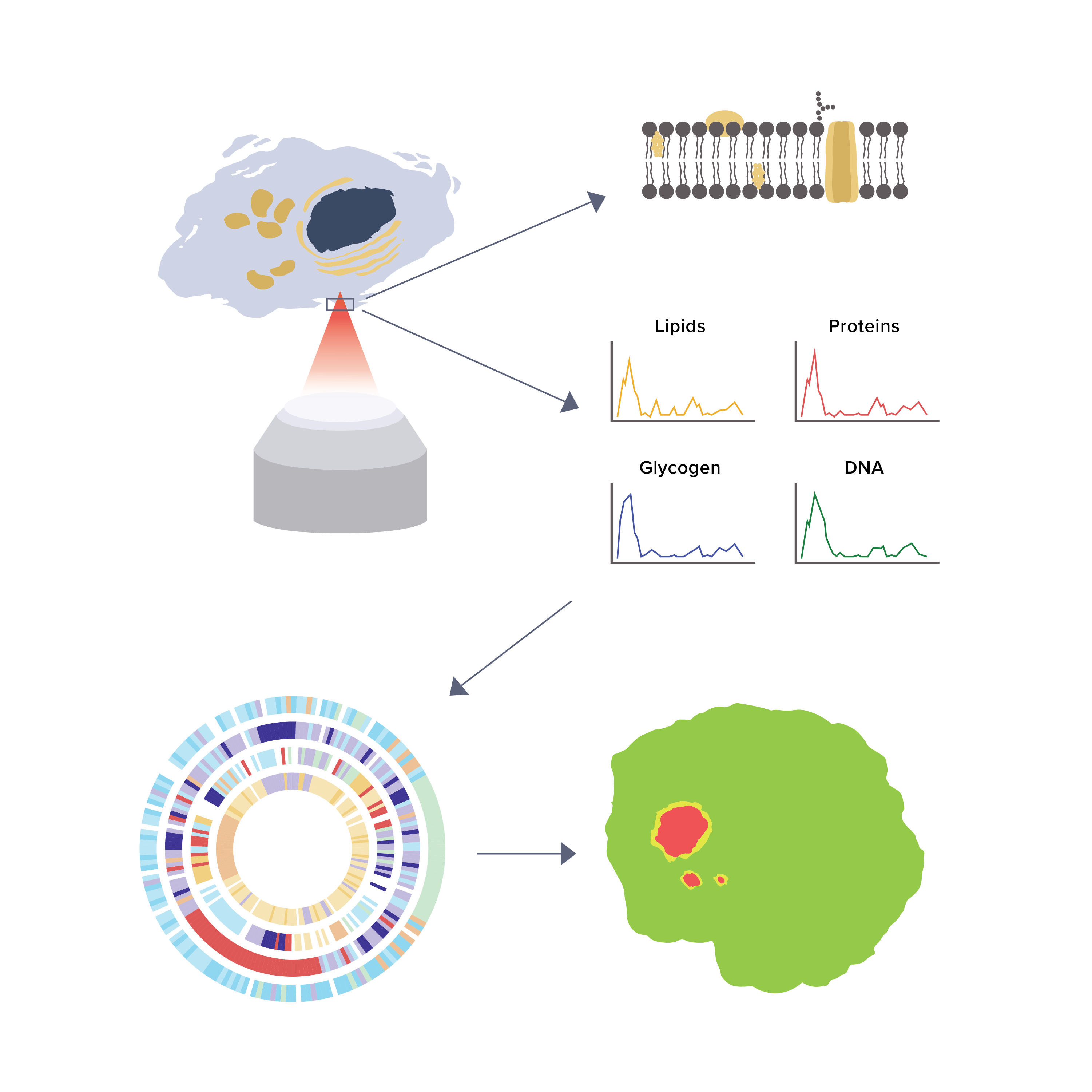
Lipidomic Analysis using Raman Spectroscopy at Single-Cell Resolution
Spontaneous Raman spectroscopy techniques can acquire the full complement of biomolecular vibrational information and identify a wealth of biomolecules (i.e., specific biomolecular structures and conformations of proteins, lipids, nucleic acids, etc.) in tissue, without labeling, using endogenous molecules as a contrast mechanism. Raman spectrum of a whole cell (and tissue) can get complicated by overlapping spectral contributions from carbohydrates, lipids, proteins, and DNA. Therefore, extracting important biological information from the Raman spectroscopy data is not trivial. We use unbiased principal component analysis (PCA) and targeted approaches using lipid standards to identify lipid classes. We capture the subtle differences in the spectra due to differences in lipid concentration and lipid classes by reducing the dimension of complex data sets using multivariate statistical methods.
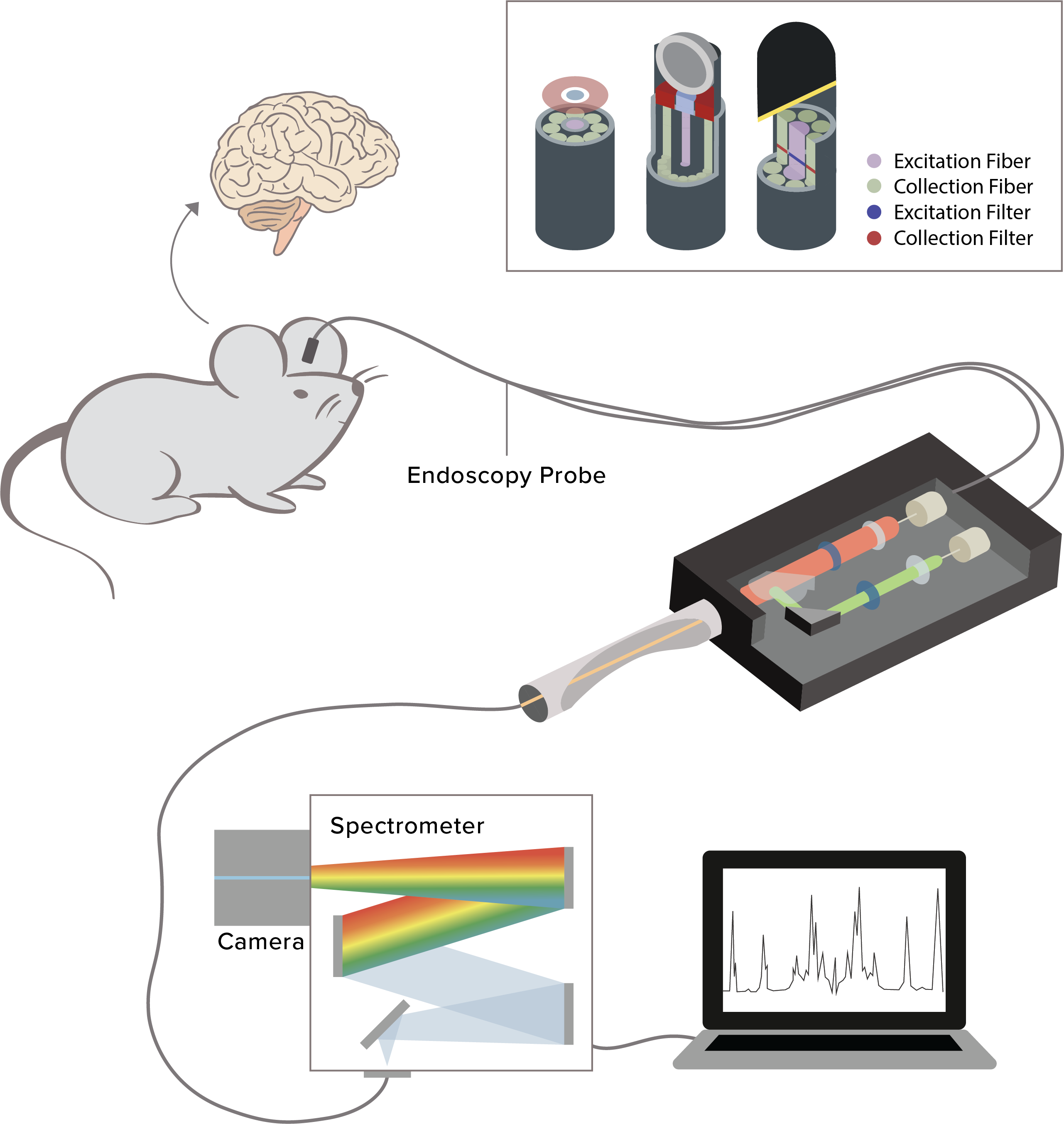
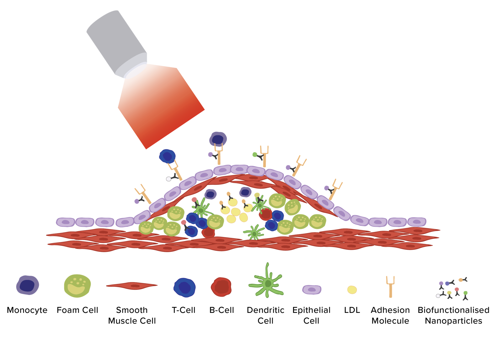
Raman Endoscopy Probe for in vivo Biochemical Analysis
The Raman fiber optics probe is designed such that it allow different spectroscopic techniques such as Raman, fluorescence, and diffuse reflectance measurements (additionally fluorescence lifetime imaging (FLIM) can be added) to be performed using the same probe. The outside diameter of the probe is 2.1 mm (0.083 in), fully immersible in most powders and aqueous solution, operating temperature range up to 15⁰ - 70⁰ C (59⁰ - 158⁰ F), excitation wavelength = 785 nm (capability to add either 633 nm or 830 nm), and maximum input power = 100 mW. The excitation light is delivered through a single co-axial optical fiber (300 µm core) and the Raman scattered light is collected using seven 300 µm core fibers. The illumination spot size of the system is 500 µm.
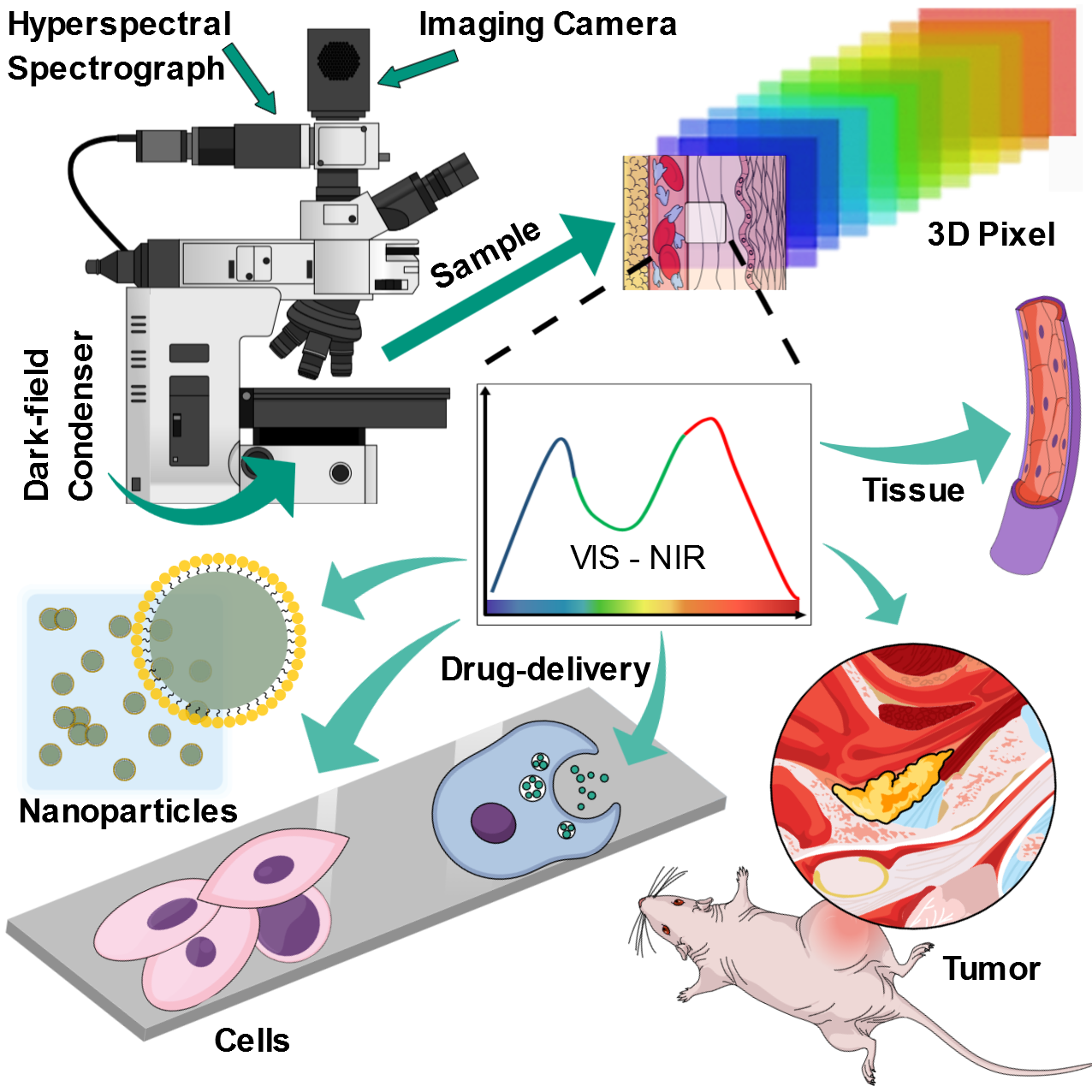
Hyperspectral Imaging
Dark-field hyperspectral imaging (HSI) combines spectroscopy and microscopy to investigate cellular transport of nanoparticles, biochemical imaging of microorganisms, single cell, and proteins. Our HSI system operates between 400-1000 nm, spectral resolution 1.5 nm, automated stage with 10 nm resolution, uses quartz Halogen Aluminum Reflector as light source, and ENVI 4.8 image analysis software. We demonstrated HSI data combined with spectral angle mapping (SAM)-based machine learning analysis to distinguish differentiating human adipose-derived stem cells (hASCs) from control stem cells. Further applications include label-free imaging of cancer cells and tissues using their endogenous optical biomarkers.
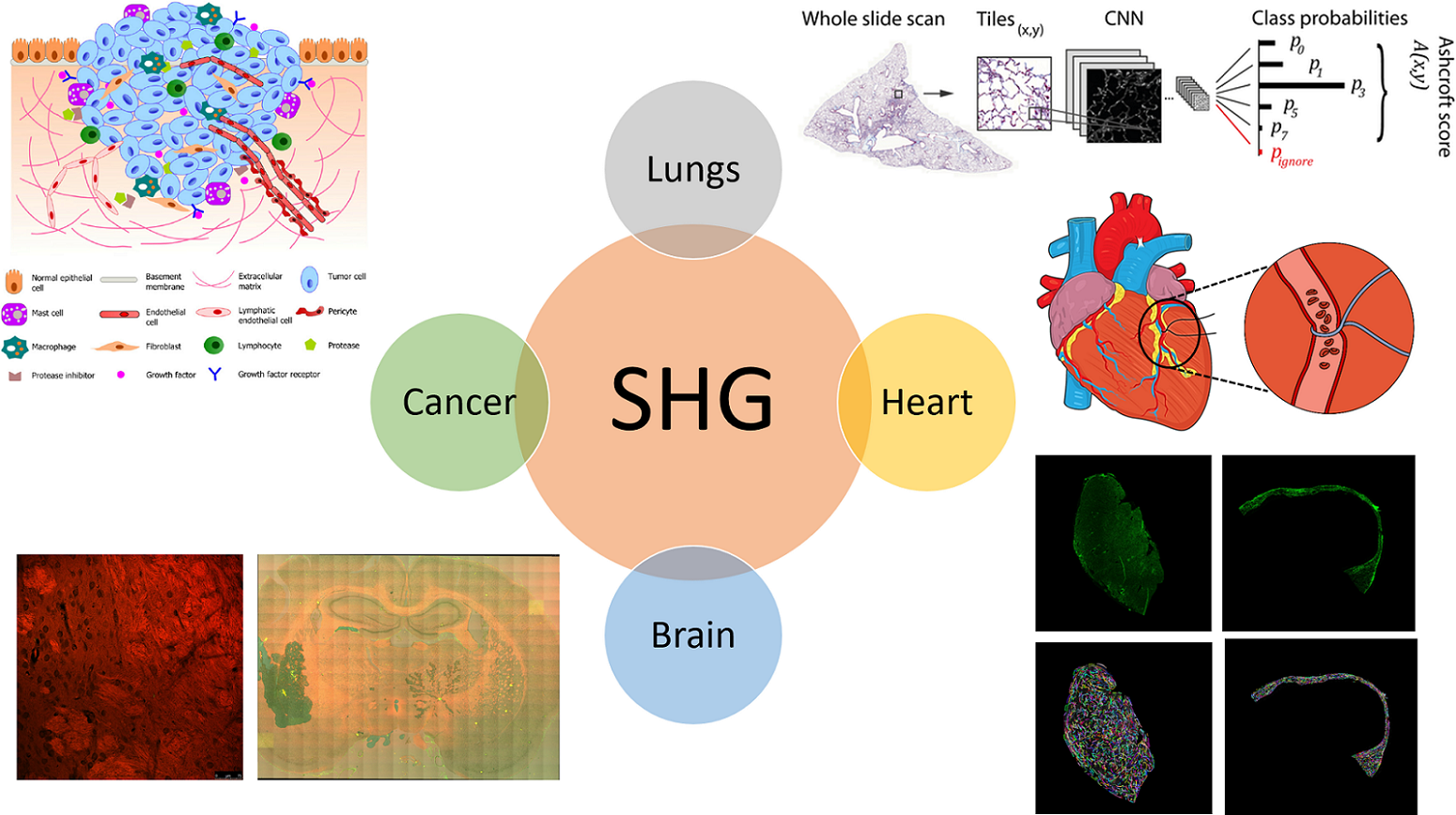
Second Harmonic Generation (SHG) Microscopy
We are using SHG imaging to investigate fibrosis in cardiac and lungs tissue. We have utilized SHG microscopy to probe the interaction between small molecule (e.g. drugs) and lipid bilayer of cells. Further studies include probing cancer microenvironment and extracellular matrix (ECM), metabolic imaging in stem cells and tissues using SHG.
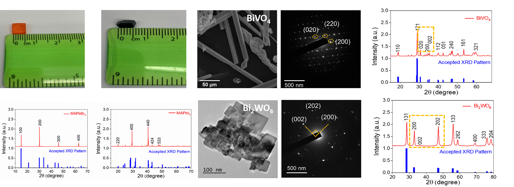
Perovskite Single Crystals for Gamma ray Spectroscopy and Solar Cells
We have synthesized several lead halide perovskite single crystal materials, including CsPbX3 (where X= Cl, Br, I), and inorganic-organic perovskites such as methylammonium lead halide, (MAPbBr3, MAPbI3). We are using these materials to make devices for gamma spectroscopy and solar cell applications. Other nanomaterials we are working with include BiVO4 and Bi2WO6. We are exploring the anti-bacterial and cancer nanomedicine properties of these materials.
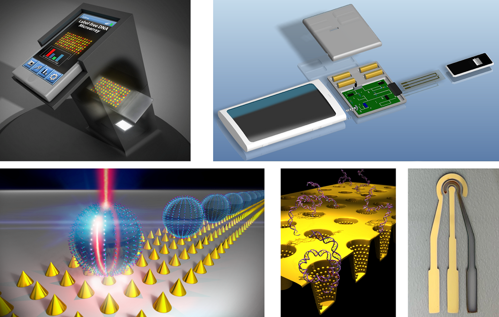
Biosensors and Smartphone Integrated System
We perform both basic science and applied research to understand, design, and manufacture sensors for biomedical, energy, forensics, and environmental applications. Some examples of sensors we have worked in our lab includes plasmonic sensors to detect cancer biomarkers, smartphone integrated electrochemical sensors to detect nitrate and phosphate in environmental water, and paper-microfluidic sensors for breast cancer genes.