Area of Interest
The recent advances in X-ray and neutron sources, detectors, mathematics, and computation
now make possible the practical use of tomography for chemical and material science
investigations. A search of NSF awards (tomography, CHE) shows that we have the first
and largest award for tomography for chemical applications, an NSF GOALI (grant opportunities
for academic liaison with industry). The collaborative partner is Dr. Larry Simeral
at Albemarle Corporation.
Dr. Kyungmin Ham, postdoc and now staff scientist at CAMD, built the first tomography
beamline at the LSU CAMD synchrotron. As one of only eight synchrotrons in the USA,
it gives us a powerful advantage for tomography research. For example, we learned
of a new tomography mathematical insight in a June, 2005 meeting; we re-programmed
the tomography beamline system by August to use this new data acquisition scheme.
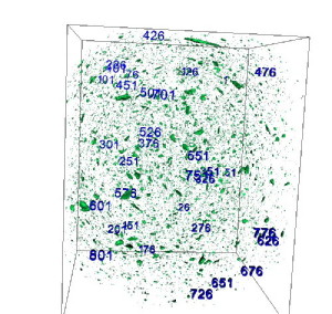
One current research project is the heat-induced dissolution of flame retardant particles
in a polymer blend. Shown here is an X-ray tomography data set rendered as an isosurface
at a threshold set to show undissolved flame retardant particles from a sample intentionally
prepared far from equilibrium. As this sample is heated, the flame retardant particles
dissolve into the polystyrene. Here, over 1400 particles are tracked across the heating
process. Each particle is analyzed with respect to volume, shape, and local concentration
gradient. The static shape analysis is from a spherical harmonic analysis procedure
developed by Dr. Ed Garboczi (NIST). We added to his code the ability to extract concentration
gradients around each tracked particle. This and other new procedures developed in
our lab will advance 3D+ materials analyses. The image was created by undergraduate
Roshan Pandrey, calculations by REU student Kameron Kilchrist, and data set collection
was done with Kyungmin Ham at the APS BM-2 beamline.
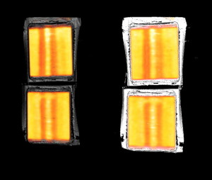
Lithium-ion polymer batteries are extremely interesting topics for 3D+ imaging. A
standard research approach would be to build a laboratory battery optimized for the
tomography experiment. However, our colleagues in the battery industry express doubts
about the relevance of this approach. A more challenging, but fruitful experimental
design is to image commercial batteries, developing new imaging procedures as needed.
Shown here are a pair of single-cell, 145 mAhr batteries with graphite/cobalt oxide
electrodes. The 3D+ neutron imaging was done so as to enhance the visibility of the
LiC6 species in the graphite electrode, shown here as the difference image between
charged and discharged batteries (left and right images show two different viewing
options). The vertical structure is attributed to volume changes in the battery as
it discharges. The images were made by REU student Bradley Wood and the data collected
at FRM II (Munich).
Our imaging work is done at a variety of sites:
- LSU CAMD synchrotron tomography beamline.
- APS BM-2, BM-32, BM13
- NIST neutron imaging beamline
- FRM II (Munich) neutron imaging beamline
- Paul Scherrer Institute (Switzerland) neutron imaging beamline
- We are involved in several construction projects for new beamlines:
- ORNL SNS VENUS neutron imaging (core member of the instrument development team)
- LSU laboratory X-ray interferometry/tomography system (our own design)
Testing Keynote posting