Research
Soft Matter and Neutronscattering
We are trying to understand the beautiful world of soft matter by a variety of techniques. At present, we concentrate on polymer melts, polymers in solution that may form micelles or vesicles. In virtually all of these cases, internal interfaces determine the material behavior.
Our focus is on fundamental science with a mission to accomplish a broader societal impact, by trying to understand materials based on our results. We target mostly model materials synthesized in our laboratory or by our collaborators. We utilize fast field cycling NMR, dielectric spectroscopy and rheology to access the material behavior often called macroscopic properties. In many cases, understanding of these results requires the knowledge of the structure and dynamics at the nanoscale. Neutron and X ray scattering techniques are our most important tools to explore the structure in the region from 0.1 to 1000 nm, and the dynamics in a window from ps to several hundred ns. Typically we utilize small-angle neutron scattering and small-angle X ray scattering, neutron spin echo and quasi elastic neutron spectroscopy. Often, we complement scattering studies by transmission electron microscopy to advance development of models to analyze scattering data.
1 Synthesis

Figure 1. Vacuum synthesis line of our laboratory. We can reduce the pressure down to 10-7 mBar.
2 Local Characterization Techniques
Our most important techniques are dynamic light scattering, fast field cycling NMR, dielectric spectroscopy, rheology, transmission electron microscopy. Our group strongly contributes to our small-angle X ray scattering instrument at the synchtrotron of the Louisiana State University, the Center for Advanced Microstructures and Devices (CAMD).
2.1 Unified description of the viscoelastic and dielectric global chain motion…
… or chasing the Holy Grail of polymer science (Macromolecules 2011, 44, 7430–7437). Once knowing the microscopic dynamics of polymers chains, as revealed by neutrons the macroscopic properties can be calculated by one theory without any assumption. The following plots show the (a) mechanical and (b) the dielectrical properties of polymer chains. The full lines show the theoretical description, by one single theory without free parameters.

Figure 2. (top) Rheology data and (bottom) dielectric spectra on polyisoprene with different molecular weights, represented by the number of entanglements Z. The lines illustrate the theoretical description.
Figure 2 a illustrates the loss modulus of entangled polymer melts obtained by oscillatory rheology and Figure 2b the dielectric loss of the same entangled polymer melts measured by broadband dielectric spectroscopy.
The lines depict a joint description of the rheology and dielectric data sets based on the tube model.
The result illustrates the insensitivity of dielectric spectroscopy to the so-called constraint release, which permits that reptation and contour length fluctuations can be well analyzed.
Constraint release has a substantial contribution to the rheology spectra. Using a combined analysis reduces the number of assumptions, and permit to identify the contribution of constraint release very precisely.
2.2 Power of Combining Dielectric Spectroscopy and Fast Field Cycling NMR
 Figure 3. Susceptibility as a function of frequency from fast field cycling relaxometry
at various temperatures.
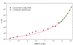
Figure 4 Relaxation time as a function of the reciprocal temperature. Fast field cycling and dielectric spectroscopy can capture ~ 12 orders over magnitude in time.
2.3 Microscopic origin of the terminal relaxation time of polymers in nanocomposites
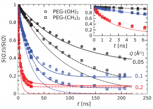
 Figure 5. (a) Dynamic correlation function S(Q,t), normalized to the static structure
factor S(Q) as a function of time t, by neutron spin echo spectroscopy. (inset) Change
of tube diameter due to adding nanoparticles. (b) Lossmodulus G" as a function of
frequency by rheology.
Figure 5. (a) Dynamic correlation function S(Q,t), normalized to the static structure
factor S(Q) as a function of time t, by neutron spin echo spectroscopy. (inset) Change
of tube diameter due to adding nanoparticles. (b) Lossmodulus G" as a function of
frequency by rheology.4. Characterization techniques at large-scale facilities
4.1 Neutron Spin Echo Spectroscopy to Understand the Dynamics in Nanocomposites
 Figure 6. Neutron Spin Echo Spectroscopy on polymers adsorbed with backbone and chain
ends.
Figure 6. Neutron Spin Echo Spectroscopy on polymers adsorbed with backbone and chain
ends.4.2 Small-Angle Neutron Scattering and In Situ Sonochemistry
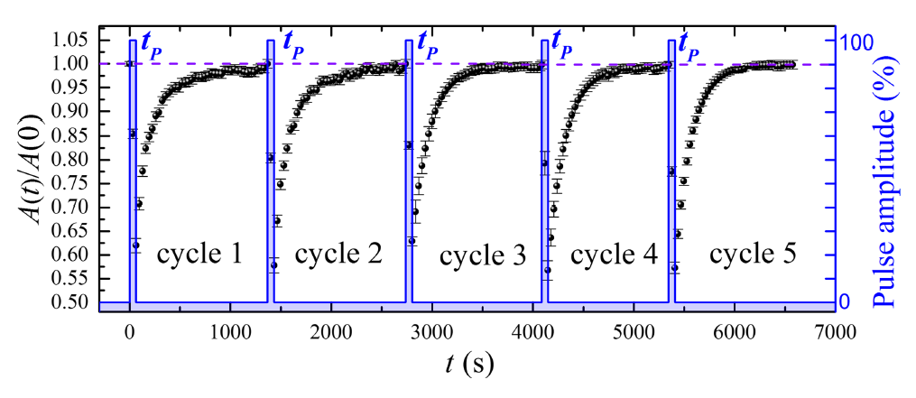
 Figure 7. Recently we introduced sonochemistry at a small-angle neutron scattering
instrument. Using this novel tools, unprecedented experiments can be conducted.