Research
Morphological innovations allow organisms to adapt to new niches and exploit new ecological opportunities, yet how such innovations arise has been a longstanding problem in evolutionary biology. A mechanistic understanding of the cellular and molecular bases of morphological innovations relies on organismal models amenable to experiments in a laboratory setting. In my laboratory we exploit the fascinating four-eyed fish Anableps anableps as a model for investigating innovations of the visual system. Anableps inhabits the waterline and is capable of simultaneous above and below water vision. Light from above or below the waterline enters the eye through uniquely duplicated set of corneas, and traverses a single pear-shaped lens, to finally reach the retina. The simultaneous aerial and aquatic sensory information is relayed to still-uncertain retinorecipient areas in the brain. My lab seeks to uncover the genetic and developmental mechanisms behind this evolutionary innovation, offering insights into the evolution of vision and the brain areas processing visual information using the zebrafish and the four eyed fish as model systems.
Projects
Partial duplication of the eye
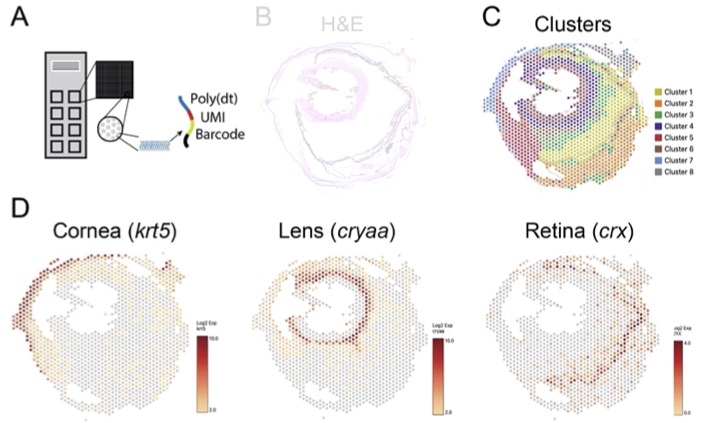
There are many intriguing questions about adaptation and diversification that evolutionary developmental biology can answer, but they aren't always easily accessible. When an embryo is developing, there's a genetic blueprint that tells the body how to form all its organs, including the eyes. This process is conserved in many animals, such as humans, birds, and other fish. However, in the four-eyed fish, something unusual happens that causes the cornea and pupils to duplicate. This species is a prime example of nature's innovation of the visual system , making them an excellent model organism to study evolutionary novelty. To better understand the process of cornea and pupil duplication, we use cutting-edge spatial RNA-seq to identify candidate genes showing expanded, dorsalized or ventralized expression patterns during cornea duplication.

Neurocranium adaptations
The ability to see above and below the water enhances A. anableps prey acquisition
and allows them to exploit aquatic and terrestrial prey populations. In this species,
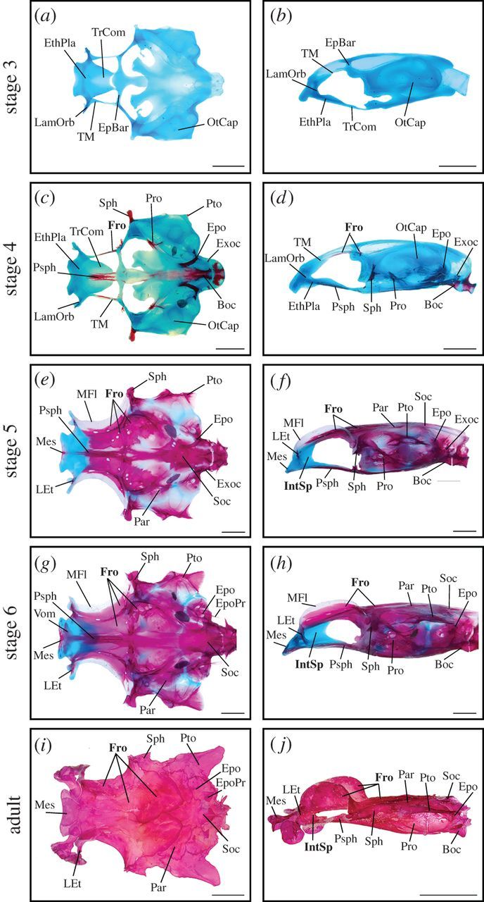
a series of unique neurocranium modifications accompany the development of its enlarged
eye. The Anableps eye is surrounded by an ossified interorbital septum dividing the
orbital cavities; through ontogenetic analysis of the neurocranium, we observed that
the frontal bone expansion coincides with the development of the eye. These observations
indicate that changes to the neurocranium and eye morphology are developmentally linked,
and we aim at understanding the mechanisms underpinning the expansion of these structures
in the neurocranium using micro CT, C&S, RNA-seq and LoF.

Gene regulatory variation and retina sub functionalization
During vertebrate eye development, the optic vesicle invaginates and forms the optic
cup, which later gives rise to the retina. The presumptive retina then becomes spatially
regionalized into a dorsal domain marked by expression of Tbx5, and a ventral domain
characterized by the expression of Vax2. The gene cascade of retina DV patterning
is well studied, yet little is known of the cis-regulatory landscape coordinating
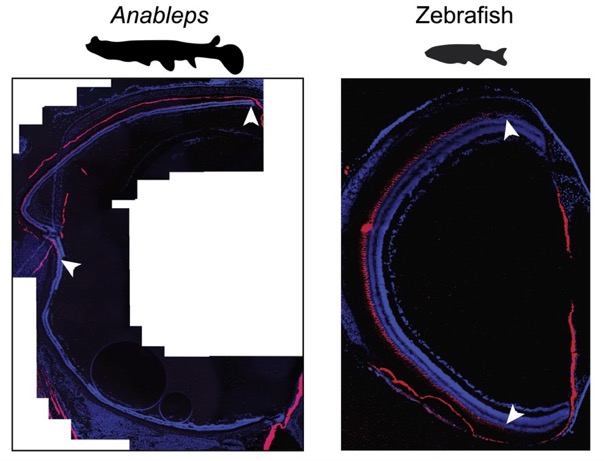
gene expression in these spatial domains. Contrary to teleosts such as the zebrafish,
the adult Anableps retina has dorsal and ventral domains that occupy each approximately
half of the retina, show distinct morphology and unique opsin gene expression profiles.
We use the Anableps retina to identify CREs that maintain the retina gene expression
territories combining ATAC-seq, Spatian RNA-seq and zebrafish transgenesis.

Mapping the retinorecipient areas in the brain
In fish, the visual input is collected in the ganglion cell layer and projected from the retina through the optic tract, crossing the chiasma and entering the brain in the contralateral side, ultimately innervating the diencephalic optic tectum. The conversion of visual sensory information into motor reactions requires specialized neural processing, frequently spread across various retinorecipient brain structures. These retinorecipient areas have been described in detail in zebrafish and are called arborization fields (AFs). The Anableps projects to the brain a single optic nerve from each eye. however, retina receives simultaneous aquatic and aerial information we want to address whether there are coevolving traits enabling the phenotypic integration of the duplicated eye structures as morphological innovations. We use use neuronal labeling, microCT and antibody staining to map the areas receiving visual input.
